New paper: a quick stain to see symbionts inside whole animals
How do you check whether an animal still carries its symbiotic bacteria without cutting it into histological sections? Together with Valentin Kokarev, Kiana Alfaro and Suzanne Dufour (Memorial University of Newfoundland), we tested SYBR stains (dyes normally used to stain DNA in agarose gels) for confocal imaging of whole chemosymbiotic animals.
SYBR Gold and SYBR Safe revealed the symbionts in the gills of thyasirid clams and inside the trophosome of the tubeworm Siboglinum fiordicum. The stains are cheap, bright, and need only the standard 488 nm laser.
The method allowed us for the first time visualize that in male Siboglinum the symbiont-housing organ is not restricted to the rear of the body, as previously described, but extends forward and wraps around the testes. We now plan to use this stain to screen our laboratory cultures and to follow how larvae acquire their symbionts.
Kokarev V.†, Rimskaya-Korsakova N.†, Alfaro K.M., Dufour S.C. (2026). Micron 208, 104120. Open access: doi.org/10.1016/j.micron.2026.104120
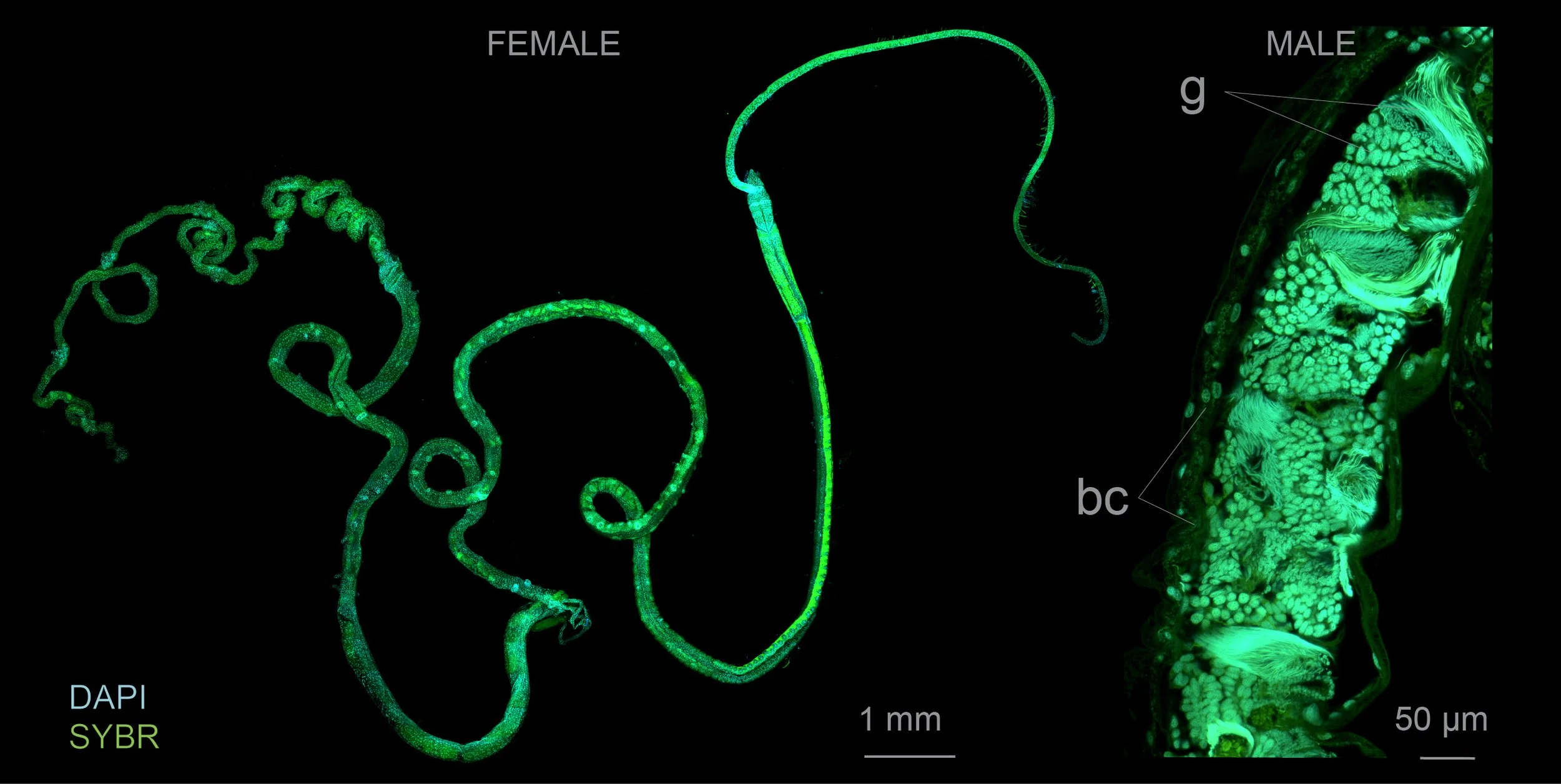
Whole mounts of Siboglinum fiordicum stained with SYBR Safe (green) and DAPI (cyan). Left: female — overview. Right: male — the trophosome (bc) surrounds the developing gametes (g). bc, bacteriocytes; g, gametes. From Kokarev, Rimskaya-Korsakova et al. 2026, Micron (CC BY 4.0).